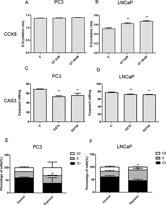
Figure 4. Oxytocin (5 nmol/L; 50 nmol/L) increases the proliferation of LNCaP cells and reduces the apoptosis of the PC3 and LNCaP cells.

(A, B) Oxytocin elevates the proliferation of LNCaP and no significant difference is observed in the PC3 cell line. (C, D) Oxytocin reduces apoptosis of the LNCaP and PC3 cell lines in both doses. (E) In the PC-3 cell line, oxytocin increase the average proportion of S phase from 24.68 ± 0.26% to 29.85 ± 1.88% (p < 0.05). (F) In the LNCaP cell line, oxytocin tends to decrease the average proportion of G0/G1 phaseand and increase the average proportion of S phase from 21.72 ± 3.45% to 42.09 ± 5.43% (p < 0.01) significantly. All The values are shown as the means ± SD. *P <0.05, **P < 0.01 (compared with the control group); N = 3-5. Abbreviations: C: control, OT: oxytocin, CAS3: caspase3.
