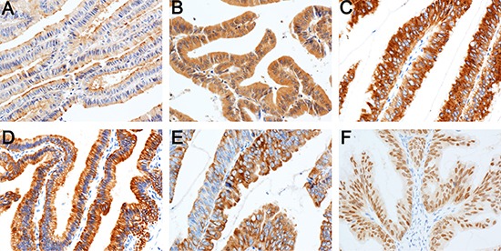
Figure 2. Expression of immunohistochemical markers in tumor cells.

MUC1 in the apical membrane (A) and cytoplasm of tumor cells (B) MUC2 (C) CK7 (D) and CK20 (E) in the cytoplasm; CDX2 in the nucleus (F). Immunohistochemical staining 200×.

MUC1 in the apical membrane (A) and cytoplasm of tumor cells (B) MUC2 (C) CK7 (D) and CK20 (E) in the cytoplasm; CDX2 in the nucleus (F). Immunohistochemical staining 200×.