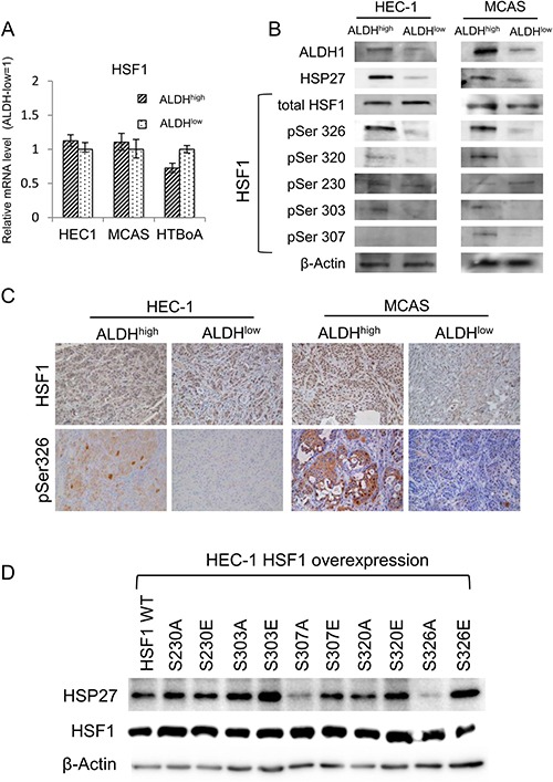
Figure 4. Stress-activated transcription factor HSF1 works upstream of HSP27.

(A) HSF1 expression by qRT-PCR analysis. HSF1 expression was assessed by qRT-PCR using ALDH1high and ALDH1low cells derived from HEC-1, MCAS and HTBoA cells. GAPDH was used as an internal control. (B) Western blotting analysis. ALDH1high cells and ALDH1low cells in HEC-1 and MCAS cells were used. The expression of ALDH1, HSP27, HSF1 and phospho-HSF1 (pSer326, pSer320, pSer230, pSer303 and pSer307) was evaluated by Western blotting. β-actin was used as an internal control. (C) Immunohistochemical findings of Ser326-phosphorylated HSF1. Tumors derived from ALDH1high cells and ALDH1low cells of HEC-1 and MCAS cells were immunohistochemically stained using anti-phospho-HSF1 (pSer326) antibody and anti-HSF1 antibody. Magnification ×200. (D) Expressions of HSP27 in HSF1 mutants overexpressed HEC-1 cells. HEC-1 cells were overexpressed with HSF1 wild type (WT) and mutants (S230A, S230E, S303A, S303E, S307A, S307E, S320A, S320E, S326A and S326E). The expression of HSP27 protein and total HSF1 were analyzed by Western blots. β-actin was used as an internal control.
