
Figure 5. Niclosamide suppresses tumorigenic potential and EMT in vivo.
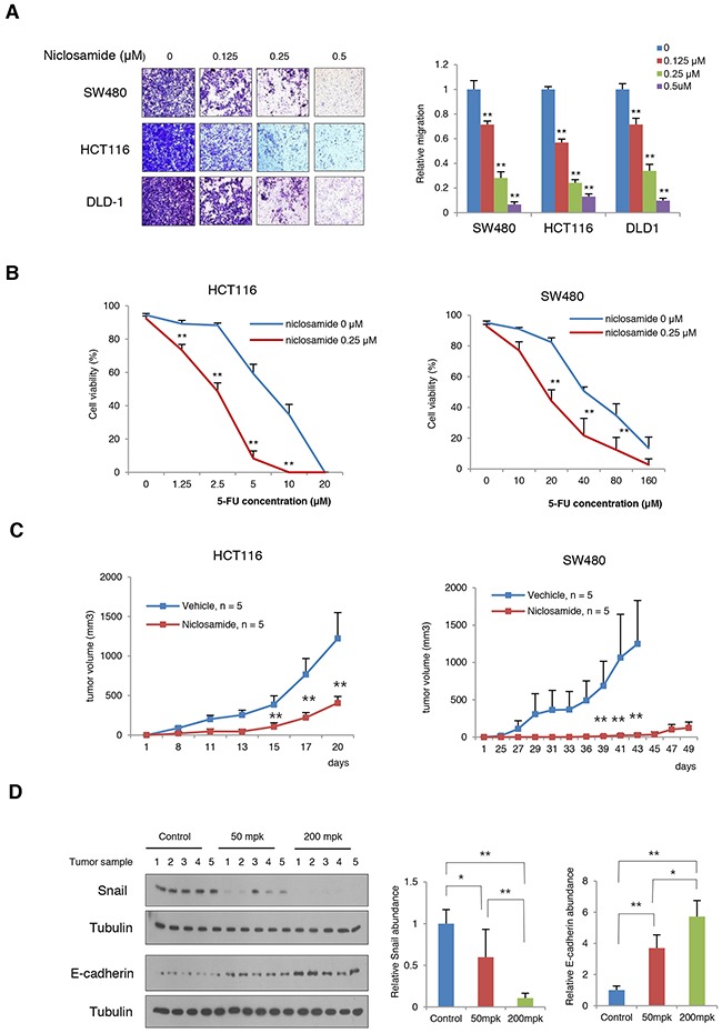
(A) The migratory activity of colon cancer cells treated with various concentrations of niclosamide was determined by transwell migration assay (left panels). Statistical significances compared to control (right panel) are denoted as **, P < 0.01 by a two-tailed Student's t-test. (B) Combination effect of niclosamide and 5-FU on HCT116 and SW480 cells. The colon cancer cells were treated with various concentrations of 5-FU and niclosamide as indicated. Cell viability was measured trypan blue exclusion assay after 48 h of incubation. **, P < 0.01 by a two-tailed Student's t-test. (C) Niclosamide inhibits the growth of colon cancer cells in vivo. HCT116 and SW480 colon cancer cells were inoculated into the flank of athymic nude mice prior to 24 h of treatment. Niclosamide (50 mg/kg dissolved in Cremophor EL) was intraperitoneally administered. Two asterisks, P < 0.01 compared to the vehicle control by Mann-Whitney test. (D) Niclosamide treatment reversed Snail-mediated EMT in SW480 xenografts. Tumor-bearing nude mice were given 50 mg/kg or 200 mg/kg body weight niclosamide for 3 days prior to sacrifice. Tumor tissues were collected and the protein abundances of Snail and E-cadherin in tumor lysates were then measured using immunoblot assay (left panels). The band intensity on each blot was normalized to the loading control and the relative protein abundance is shown as the ratio to that in the vehicle control (middle and right panels). Results are shown as means and s.d. One asterisk, P < 0.01; Two asterisks, P < 0.001 compared to the vehicle control by Mann-Whitney test.