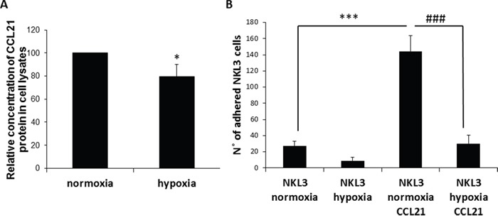
Figure 2. Influence of CCL21 presentation by peripheral lymph node endothelial cells and hypoxia on NK cells recognition and adhesion.

A. Hypoxia vs normoxia CCL21 production by HPLNEC.B3 cells measured by ELISA in cell lysates. Concentration of CCL21 protein produced under normoxia was set as 100%. Values marked with a star vary significantly (p<0.05, N=3). B. Quantification of the NKL3 adhesion to HPLNEC.B3 as number of NKL3 cells counted on the surface of HPLNEC.B3 cells (ten representative fields were counted) in normoxia and in hypoxia showing a reduction before as well as after treatment with CCL21. NK cells were labelled by the PKH26 red fluorescent cell linker kit. NK cells were injected on the HPLNEC.B3 cells monolayer (1.106 NK cells/ml) at a fixed flow rate of 0, 2 dyn/cm2 for 5 minutes and washing by OptiMEM at 0, 6 dyn/cm2 for 10 min. Adhered NK cells amount was quantified. *** p<0.001, N=3, ### p<0.001, N=3.
