Figure 4.

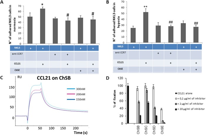
A. Impact of CCL21 presentation by GAGs on CCR7+ NK cells adhesion to endothelial cells. Quantification of the NKL3 adhesion to HPLNEC.B3 as number of NKL3 cells counted on the surface of HPLNEC.B3 cells (ten representative fields were counted) in normoxia (A) and in hypoxia B. showing an increase in adhered NKL3 after treatment with CCL21 and decrease after treatment with CCR7 neutralizing antibody and chondroitin sulfate in both conditions. NK cells were labelled by PKH26 red fluorescent cell linker kit then, treated or not with CCR7 neutralizing antibodies (10 μg/ml), or ChSE (0,6 μg/ml) for 1 hour at 4°C. Then, NK cells were injected on the HPLNEC.B3 cells monolayer (1.106 NK cells/ml in OptiMEM) at a fixed flow rate of 0, 2 dyn/cm2 for 5 minutes and washing by OptiMEM at 0, 6 dyn/cm2 for 10 min. Adhered NK cells amount was quantified. * p<0.05, N=3, # p<0.05, N=3; ** p<0.005, N=3, ## p<0.005, N=3. C. Plasmon resonance assessment of the affinity of CCL21 chemokine for various glycosaminoglycans. CCL21 was injected over the GAG surface in a range of concentrations (from 0 to 300 nM) to produce sensorgrams for association and dissociation phases analysis. Binding kinetics were evaluated with a 1:1 Langmuir model using the BiaEvaluation software (Biacore). D. Inhibition experiments by co-injection of CCL21 chemokine with various concentrations of GAGs on the immobilized ChSD surface. Relative inhibition was preferential for chondroitin sulphate E compared to ChS B, D and C.
