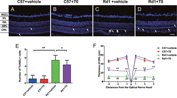
Figure 2. Effects of T0 on apoptosis and thickness of the C57/rd1 retina outer nuclear layer (ONL) at P14.

The TUNEL assay was performed at P14. (A-D) TUNEL-positive cells (green) are sparsely dispersed in the ONL of C57 mice but densely dispersed in the ONL of rd1 mice. DAPI staining (blue) was shown as a guide for retina layers. (E) There is no significant difference in the TUNEL-positive cells in the ONL of C57 mice treated with T0 or vehicle. However, the number of apoptotic cells is remarkably higher in the ONL of rd1 mice than in the age-matched C57 mice, which is significantly reduced by T0 treatment. (F) There is no significant difference in the thickness of the ONL of C57 mice treated with vehicle or T0, and the decrease in the thickness of the ONL of rd1 mice treated with T0 is markedly attenuated compared to that in the vehicle-treated control rd1 mice. RGCL, retinal ganglion cell layer; IPL, inner plexiform layer; INL, inner nuclear layer; OPL, outer plexiform layer; ONL, outer nuclear layer. n=4 in each group. **P<0.01, *P<0.05. Scale bar=50 μm.
