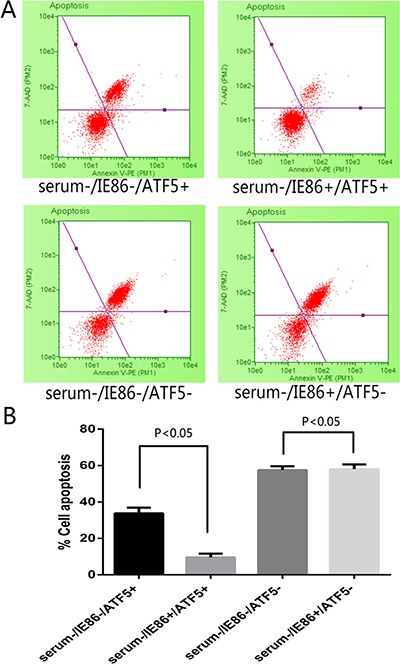
Figure 3. Apoptosis detection following treatment with serum deprivation and IE86 expression in ATF5 (+/.

−) U87 cells. (A) Apoptosis measured by Annexin V/7-AAD staining with flow cytometry after serum deprivation 72 h. Events in each of the four quadrants are as follows: Lower-left: viable cells. Lower-right: cells in the early to mid-stages of apoptosis. Upper-right: cells in the late stages of apoptosis. Upper-left: mostly nuclear debris. (B) The total apoptosis rate obtained by Annexin V/7-AAD staining after 72 h treatment with serum deprivation in different cell group. IE86-expressing cells displayed significant resistance to cell apoptosis evoked by serum deprivation, but in RNAi-ATF5 U87 cells IE could not rescue the cells from apoptosis.
