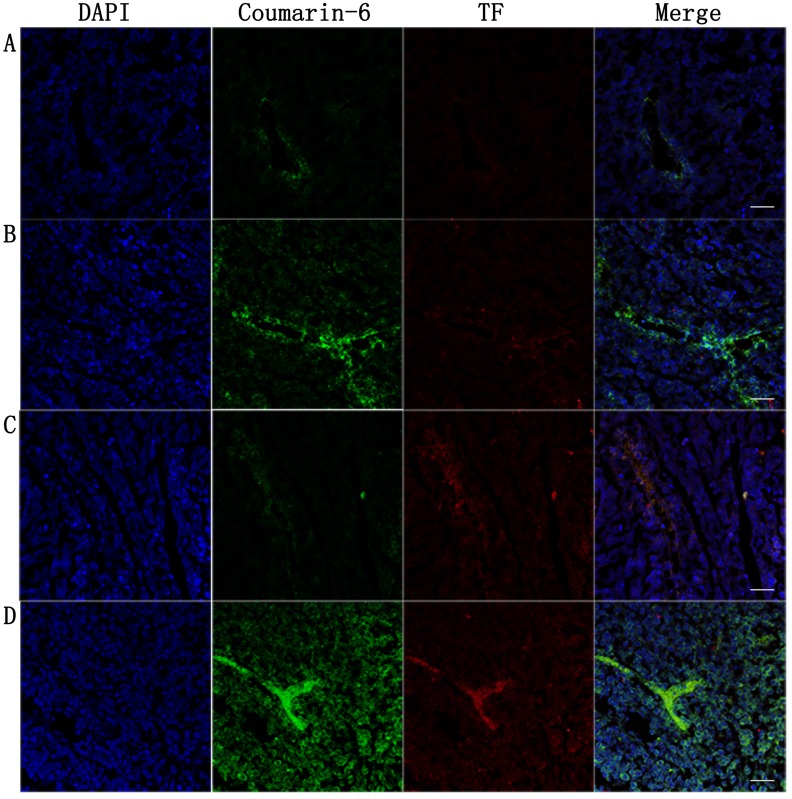
Figure 10. Tumor were harvested for insections at 24h post-PDT to estimate nanoparticles accumulation and TF expression in tumor vessels.
Tumor-bearing mice were singly administered with nanoparticlesA.,B. or combining i.v. injection of nanoparticles with PDT C.,D.. Coumarin-6-labeled NP was injected via tail vein in group A and C, and coumarin-6-labeled EGFP-EGF1-NP was injected in group B and D. Frozen sections tumors were was stained with rabbit anti rat TF polyclonal antibody examined by confocal microscopy. Blue: cell nuclei. Green: coumarin-6-labeled nanoparticles. Red: TF expression. Bar = 20um.

