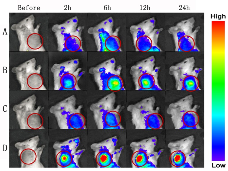
Figure 7.
In vivo multispectral fluorescent imaging of tumor-bearing mice at different time points post-PDT. Tumor-bearing mice were respectively conducted with single i.v. administration of nanoparticles A., B. and a combination of i.v. administration of nanoparticles and PDT C., D.; Dir-labeled NP was injected via tail vein in group A and C, and Dir-labeled EGFP-EGF1-NP was injected in group B and D.

