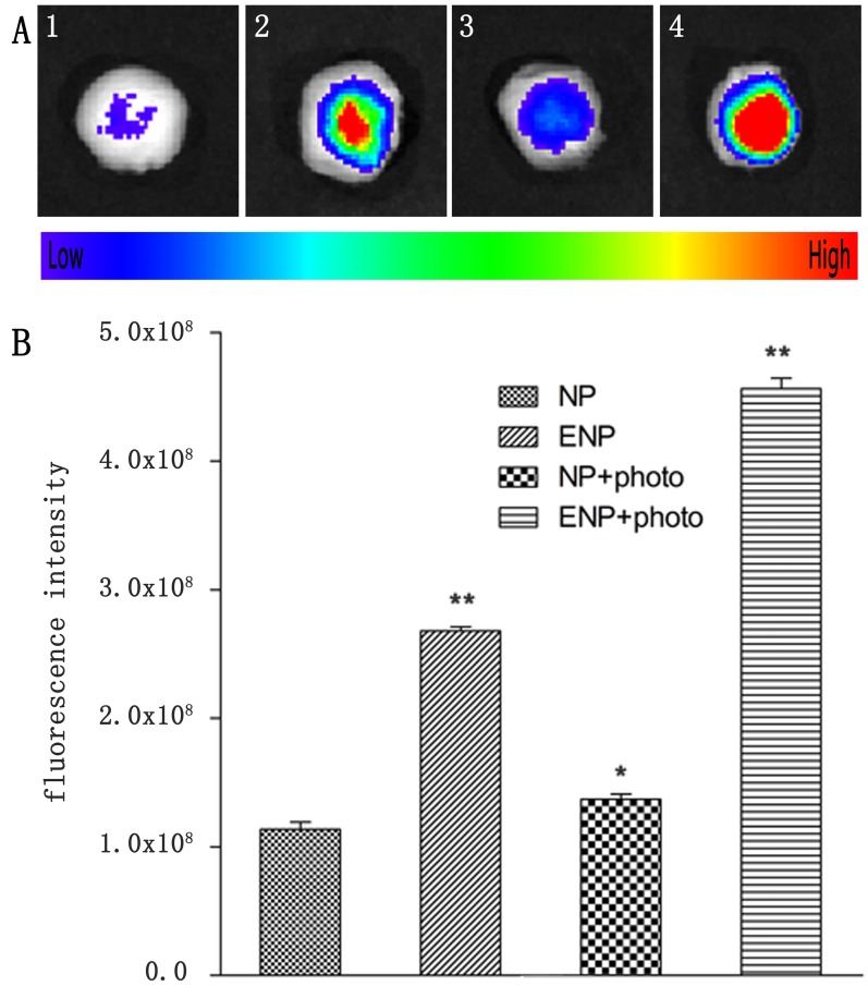
Figure 8. Tumors were incised at 24 h post-PDT for.
ex vivo multispectral fluorescent imaging. (1) single i.v. administration of Dir-labeled NP; (2) single administration of Dir-labeled EGFP-EGF1-NP; (3) a combination of i.v. administration of Dir-labeled NP and PDT; (4) a combination of i.v. administration of Dir-labeled EGFP-EGF1-NP and PDT. B. corresponding semi-quantitative fluorescence intensities of tumors. Data are expressed as mean ± SEM (n = 3); *p < 0.05, **p < 0.01, compared with tomors treated with NP.

