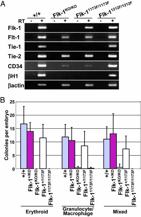
Fig. 5.
Hematopoietic defects in Flk-11173F/1173F embryos. (A) RT-PCR analysis of Flk-1, Flt-1, Tie-1, Tie-2, CD34, and βH1 with E8.5 wild-type, Flk-1KO/KO, Flk-11173F/1173F, and Flk-11212F/1212F embryos including yolk sacs. Each amplification was performed in the absence (–) or presence (+) of reverse transcriptase (RT) to detect genomic DNA contamination. β-actin was used as a positive control. (B) Hematopoietic colony formation assay of E8.5 wild-type, Flk-1+/KO, Flk-1KO/KO, Flk-1+/1173F, and Flk-11173F/1173F embryos including yolk sacs. Error bars indicate the standard deviation. Wild-type, n = 6; Flk-1+/KO, n = 8; Flk-1KO/KO, n = 6; Flk-1+/1173F, n = 8; Flk-11173F/1173F, n = 7.

