Figure 1.
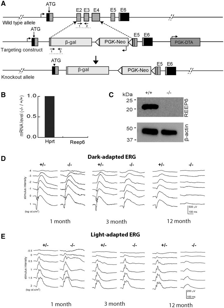
Targeted disruption of Reep6 in mice. (A) Strategy for targeting Reep6. WT locus is shown at top. Grey boxes indicate exons (1–6). Arrows below the E2/E3 exons and β-gal boxes represent the primers used for screening/genotyping. The schematic of the targeting construct is shown in the middle and the knock out locus is shown at the bottom. (B) Expression profile of mRNA transcripts collected from WT and Reep6–/– retinae, quantified by RT-qPCR using exon-specific Taqman probes. Hprt primers were used as a control. (C) Immunoblot of protein extracts from WT and Reep6–/– mouse retina (P30). The REEP6 antibody detects 23 kDa protein, which is absent in the Reep6–/– mice. (D–E) ERG traces at increasing light intensities from dark –adapted (D) and light –adapted (E) Reep6+/– heterozygous (+/−) and Reep6–/– (−/−) mice at 1 month, 3 months and 12 months of age. The amplitudes of the scotopic a –wave and the b-wave decrease significantly in Reep6–/– mice.

