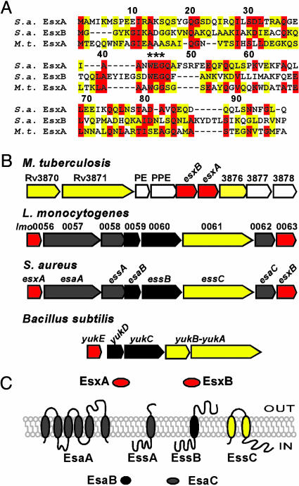
Fig. 1.
S. aureus ess locus encoding ESAT-6-like proteins. (A) Protein sequence alignment of S. aureus (S.a.) EsxA and EsxB with M. tuberculosis (M.t.) EsxA. M. tuberculosis and S. aureus EsxA display 20.8% identity and 25% similarity, whereas S. aureus EsxB and M. tuberculosis EsxA are 17.8% identical and 35% similar. All three proteins contain the WXG motif. (B) Comparison of the M. tuberculosis H37Rv ESAT-6 locus with the S. aureus, L. monocytogenes ess loci and the B. subtilis yuk locus. Color of genes and proteins indicates FSD factors (yellow), ESAT-6-like (red), mycobacterial genes (light gray), and staphylococcal (also in listeria or bacilli, dark gray). (C) Membrane topology or soluble character of proteins encoded by the S. aureus ess locus.

