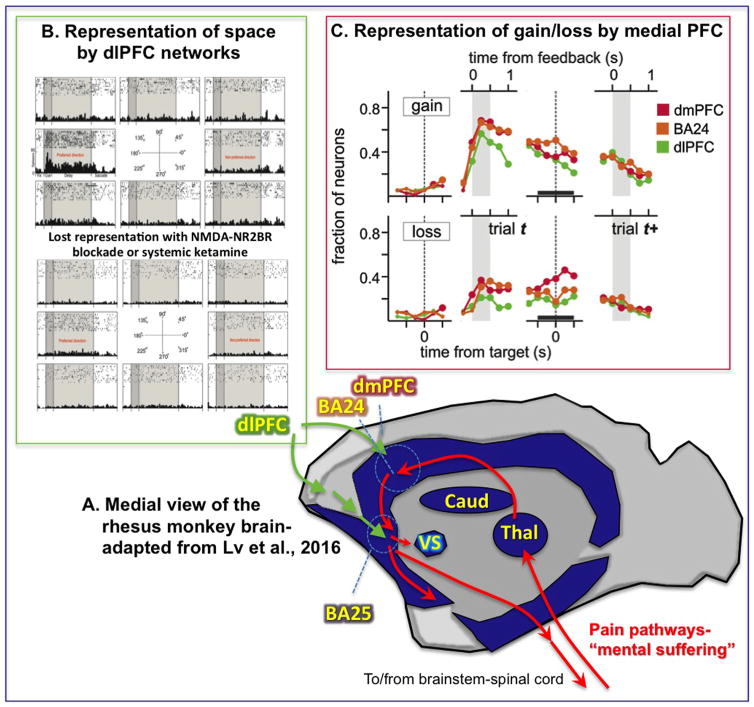
Figure 1.
(A) Schematic illustration of the results of Lv et al. (3) showing reduced functional connectivity (dark blue) in monkey medial prefrontal cortex (PFC) and subcortical structures 18 hours following ketamine. Anatomical pathways mediating the emotional aspects of pain are shown in red, with Brodmann area (BA) 24, dorsomedial PFC (dmPFC), and BA25 outlined; the dorsolateral PFC (dlPFC) (not shown, as it is on the lateral surface) connects to BA25 via rostral medial PFC. (B) Effects of N-methyl-D-aspartate (NMDA) receptor blockade on dlPFC delay cell firing, where iontophoresis of an NMDA receptor-NR2B antagonist causes complete loss of the representation of visual space. (C) Time course of neural signals related to gains and losses in dmPFC, dlPFC, and the anterior cingulate cortex (BA24). Shown are the fractions of neurons with significant gain- or loss-related activity within each region at different time lags from the time of target or feedback onset. Caud, caudate; Thal, thalamus; VS, ventral striatum. [(B) Adapted from Wang et al. (9). (C) Adapted from Seo and Lee (8)].

