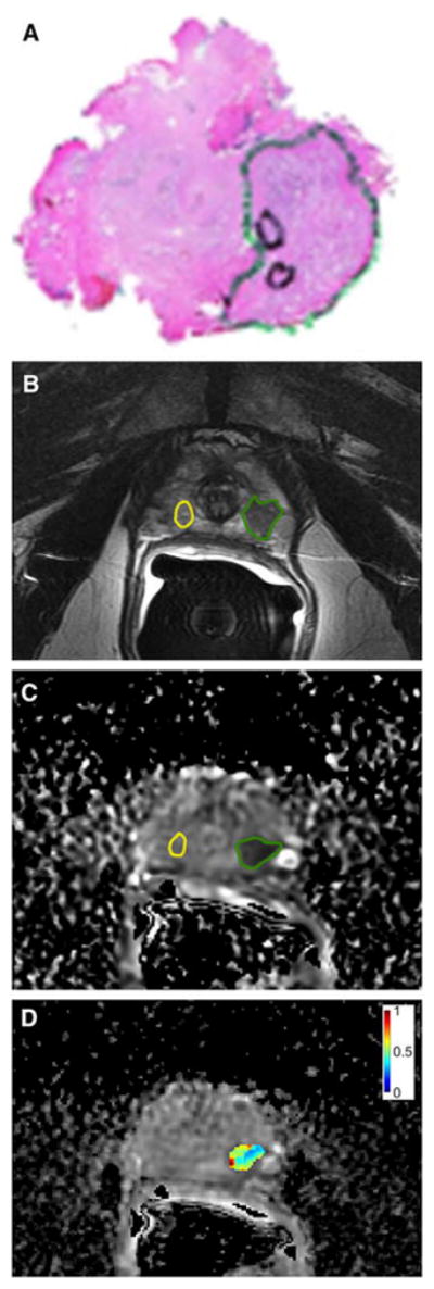
Figure 4.

Whole-mount prostatectomy histopathology specimen (a), multiparametric prostate MRI (b,c) with texture analysis (d) of a 63-year-old man. (a) Whole-mount histopathology demonstrated a Gleason 3+4 prostate cancer (green circle). (b) T2-weighted imaging showed a corresponding region of low signal intensity in the peripheral zone (green circle) as compared to non-cancerous tissue (yellow circle). (c) The cancer showed restricted diffusion on the apparent diffusion coefficient (ADC) map (green circle). (d) Texture analysis of the ADC map showing the normalized ADC Entropy map of the tumour. Reprinted with permission from [32].
