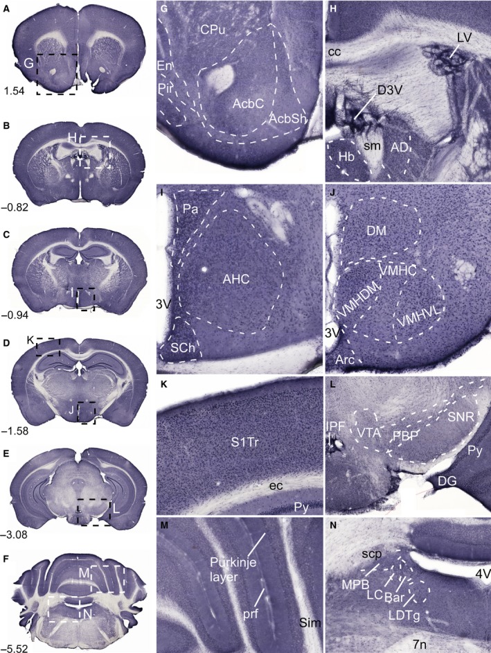
Figure 4.

Immunostaining of SLC38A10 in the mouse brain. Nonfluorescent immunohistochemistry on free‐floating sections with overview pictures (A–F) and close up pictures (G–N) of SLC38A10 immunostaining in mouse brain. (G) Staining of SLC38A10 in nucleus accumbens (Acb), caudate putamen (CPu, striatum) and in piriform cortex (Pir) (Bregma 1.54). (H) SLC38A10 immunoreactivity in cells within D3V and LV (Bregma −0.82). (I) High immunostaining of SLC38A10 in paraventricular hypothalamic nucleus (Pa), suprachiasmatic nucleus (SCh) and in anterior hypothalamic area central part (AHC) close to the 3V (Bregma −0.94). (J) Localization of SLC38A10 in dorsomedial hypothalamic nucleus (DM), arcuate hypothalamic nucleus (Arc), and in ventromedial hypothalamic nucleus (VMH) (Bregma −1.58). (K) Scattered SLC38A10 staining in cells throughout cerebral cortex (Bregma −1.58). (L) Immunostaining of SLC38A10 in ventral tegmental area (VTA), dentate gyrus (DG), and in pyramidal cell layer of the hippocampus (Py) (Bregma −3.08). (M) SLC38A10 immunoreactivity in the Purkinje layer of cells in cerebellum (Bregma −5.52). (N) Immunostaining of SLC38A10 in locus coeruleus (LC) and Barrington's nucleus (Bar) in pons close to the 4V. Additional abbreviations: endopiriform claustrum (En), accumbens nucleus core (AcbC), accumbens nucleus shell (AcbSh), corpus callosum (cc), habenular nucleus (Hb), stria medullaris (sm), anterodorsal thalamic nucleus (AD), ventromedial hypothalamic nucleus central part (VMHC), ventromedial hypothalamic nucleus dorsomedial part (VMHDM), ventromedial hypothalamic nucleus ventrolateral part (VMHVL), primary somatosensory cortex trunk region (S1Tr), external capsule (ec), interpeduncular fossa (IPF), parabrachial pigmented nucleus of the VTA (PBP), substantia nigra reticular part (SNR), primary fissure (prf), simple lobule (Sim), superior cerebellar peduncle (scp), medial parabrachial nucleus (MPB), laterodorsal tegmental nucleus (LDTg), and facial nerve (7n). The described brain regions were depicted using, The Mouse Brain, by Franklin and Paxinos 2007.
