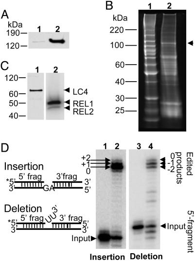
Fig. 1.
Isolation of TAP-tagged LC-2 from mitochondrial lysate of transfected L. tarentolae. (A) Equivalent amounts of cytosol (lane 1) and mitochondrial extract (lane 2) were separated on an 8–16% polyacrylamide SDS gel, which was blotted and probed with the peroxidase–antiperoxidase reagent to detect the TAP-tagged LC-2. (B) Protein composition of TAP-isolated material. Lane 1, molecular mass standards; lane 2, SYPRO Ruby stained gel (arrowhead indicates position of CBP-tagged LC-2). (C) Lane 1, Western blot probed with anti-LC-4 antiserum; the endogenous LC-4 band is indicated by an arrowhead. Lane 2, the TAP-isolated material was incubated with [α-32P]ATP before gel analysis to detect the endogenous REL1 and REL2, which are indicated by arrowheads. Note that REL2 normally labels less than REL1 as a result of being precharged with AMP (29). (D) In vitro precleaved editing activities of the TAP-isolated material. The annealed RNA substrates are shown schematically on the left. Lanes: 1, input RNA for insertion editing; 2, +2-U gRNA-mediated insertion editing; 3, input RNA for deletion editing; 4, –2-U gRNA-mediated deletion editing.

