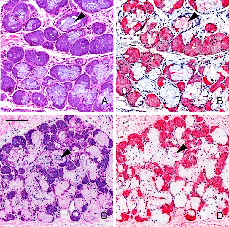
Fig. 6.
Siglec overlay of human airway submucosal glands. Cross sections of human trachea were stained with H&E (panels A,C) or with Siglec-8-Fc (B), or Siglec-9-Fc (D) precomplexed with AP-conjugated anti-human Fc. Lectin binding was detected using Vector Red stain and sections counterstained using Hematoxylin QS. Images captured using different siglec-Fc chimeras were linearly adjusted to maximize the dynamic staining range within the section. Arrowheads: selected examples of cross sections of submucosal gland ducts. Scale bar, 100 μm. This figure is available in black and white in print and in color at Glycobiology online.

