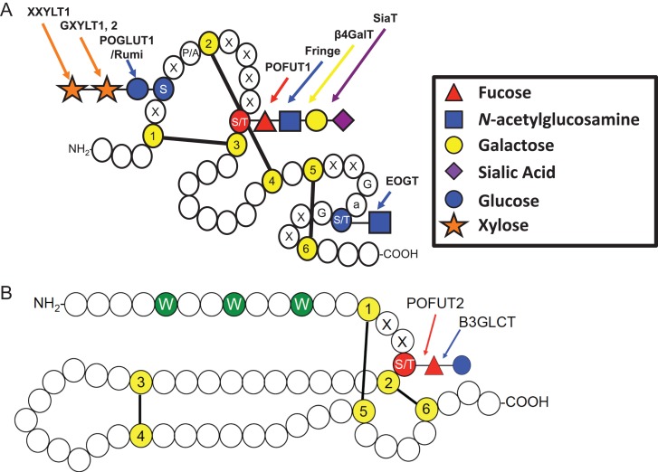
Fig. 4.
Key features of EGF repeats and TSRs. (A) Cartoon showing a single EGF repeat. Each circle represents one amino acid. Conserved cysteines (yellow) are numbered and disulfide bonds are indicated. O-Glucose and O-GlcNAc sites are shaded blue and the O-fucose site is shaded red. Enzymes responsible for the addition of each sugar are indicated. Modified from Rana and Haltiwanger (2011). Used with permission. Elsevier. (B) Cartoon showing a typical TSR. Conserved cysteines (yellow) and disulfide bonds are indicated. C-Mannose sites are shown in green and the O-fucose site is shaded red. (S) Serine; (T) Threonine; (G) Glycine; (W) Tryptophan; (X) any amino acid, (a) any aromatic amino acid. Modified with permission from Haltiwanger (2004). ©Elsevier. This figure is available in black and white in print and in color at Glycobiology online.

