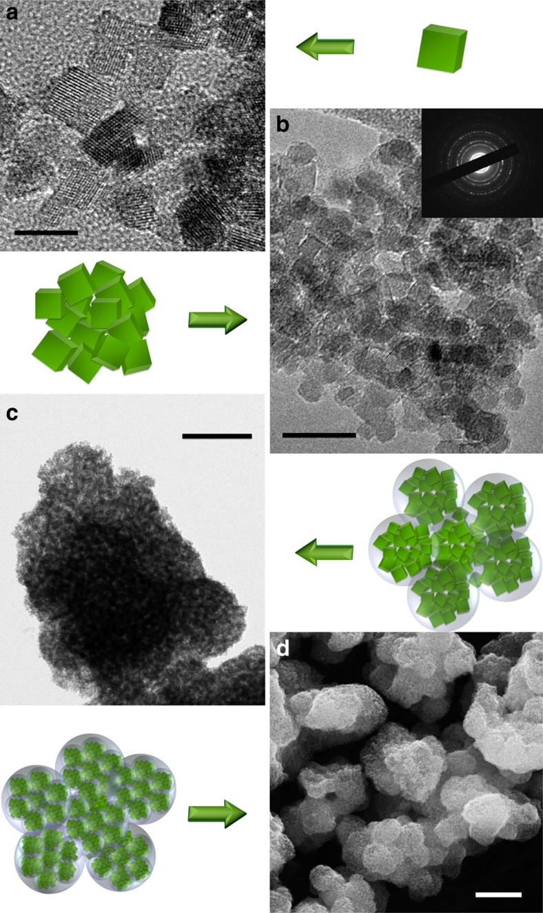
Figure 2. Hierarchical structure of the material.
(a) High-resolution TEM shows well-crystalized NPs. Scale bar, 10 nm. (b) High-magnification TEM image of single spherical aggregate. Scale bar, 20 nm. (c) TEM image of highly porous, sponge-like structure composed of spherical aggregates of fine particles. Scale bar, 500 nm. (d) SEM picture shows overall structure of spherical particles arranged into larger structures. Scale bar, 1 μm. The drawings schematically represent the observed three levels of particle arrangement with small NPs aggregated into spherical structures (150–500 nm size), which gather into even larger structures.

