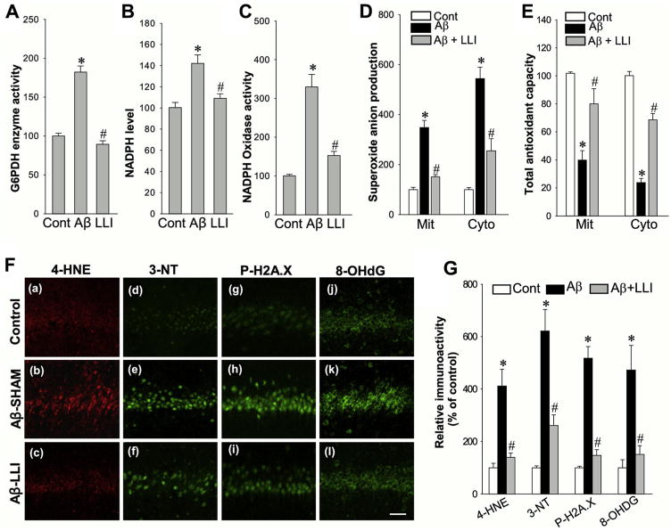
Fig. 7.
Effect of LLI on Aβ-induced G6PDH activity, NADPH oxidase activity, superoxide production and oxidative neuronal damage, and antioxidant capacity in hippocampal CA1 region. (A–E) The activities of G6PDH enzyme and NADPH oxidase, the levels of NADPH and superoxide anion production, and the antioxidant capacity were performed using the assay kits as described in detail in the Section 2. Whole-cell protein samples of hippocampal CA1 6 days after Aβ infusion were used for the detections in A–C, mitochondrial (Mit) and cytosolic (Cyto) protein samples were adopted for the assays in D and E. Data are expressed as means ± standard error (SE) from 4 to 5 animals in each group and presented as percentage changes compared with control groups. (F and G) Representative confocal microscopy images of oxidative damage markers for lipid peroxidation (4-HNE), peroxynitrite production (3-nitrotyrosine, 3-NT), DNA double-strand breaks (H2A.X Ser139), and oxidized DNA damage (8-OHdG) were taken from hippocampal CA1 region 6 days after Aβ infusion (a–l). The fluorescent intensity was quantified using ImageJ analysis software and expressed as percentage changes versus respective control group. Note that LLI strongly decreased oxidative neuronal damages 6 days after Aβ insult. Data represent means ± SE (n = 5–6 animals in each group). Scale bar: 50 μm. *p < 0.05 versus control group, #p < 0.05 versus LLI-untreated Aβ group. Abbreviations: Aβ, beta amyloid; G6PDH, glucose-6-phosphate dehydrogenase; LLI, low-level laser irradiation.

