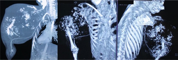
Figure 3.
CT Angiography showed extensive destruction of the osseous tissue with calcification & there appeared involvement of lateral aspect of scapula also. Blood flow in major vessels was normal without undue hyper vascularity in the tumour area & the tumour mass was pushing the thoracic cage inwards.

