Figure 4.
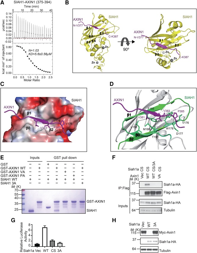
Crystal structure of AXIN1 in complex with SIAH1. (A) ITC analysis of the interaction between SIAH1-SBD and AXIN1 peptide. (B) The overall structure of the AXIN1/SIAH1 complex. The sequence of the observable part of AXIN1 in the structure is 377VPKEVRVEPQK387. (C) The surface electrostatic view between AXIN1 (377–387) and SIAH1 β0, β1, and β2 sheets is shown. (D) Major interactions between AXIN1 (377–387) and SIAH1. (E) GST pull-down analysis of the interaction between SIAH1 mutants and GST-tagged AXIN1. (F) The Siah1a 3A mutant (L176A/D177A/W178A) blocks the interaction with Axin1 in the coimmunoprecipitation assay. (G) Overexpression of Siah1a, but not the 3A mutant, enhances Wnt3a-induced STF reporter. Error bars denote the SD between four replicates. (H) Overexpression of Siah1a, but not the 3A mutant, promotes degradation of Axin1.

