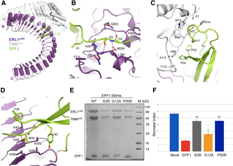
Figure 4.
Recognition mechanism of EPF1 by ERL1LRR–TMMLRR. (A) Overall structure of the EPF1–ERL1LRR–TMMLRR complex. (N) N terminus; (C) C terminus. Color codes are indicated. (B) Detailed interactions of the N-terminal side of EPF1 with ERL1LRR–TMMLRR. (C) Detailed interactions of the central region of EPF1 with ERL1LRR–TMMLRR. (D) Detailed interactions of EPF1 with ERL1LRR–TMMLRR. (E) Mutagenesis analysis of EPF1-6xHis's interaction with ERL1LRR–TMMLRR in vitro. The pull-down assays were performed as described in Figure 3A. (F) Effects of the mutations of EPF1 on the stomatal density of Arabidopsis cotyledons; the concentration of peptide was 10 µM. The error bars show SD. n > 8. (*) P < 0.05; (**) P < 0.01, versus the controls by Student's t-test.

