
Figure 1.
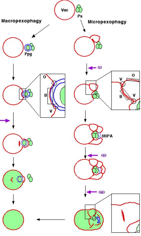
Schematic model for vacuolar membrane dynamics of two distinct pexophagic pathways and subdomains on the vacuolar membrane. Violet arrows represent possible fusion events occurring at the vacuolar membrane surface. Ps, peroxisome; Vac, vacuole; MIPA, micropexophagic apparatus; Ppg, pexophagosome; V, vertex domain; B, boundary domain; O, outside edge domain. Left: macropexophagy. A newly synthesized pexophagosome envelops a single peroxisome within a cluster, and subsequently its outer membrane fuses with vacuolar membrane. This fusion event could occur in two different ways: fusion at a contact point or fusion at a vertex. This figure represents the fusion at a vertex. Although fusion at the vertex involves internalization of the boundary region (as described in this figure), fusion at the contact point does not. Right: micropexophagy. Membrane fusion (or scission) at the vacuolar surface should occur at three different steps (indicated by violet arrows): i) septation of the vacuole; ii) completion of peroxisome engulfment; and iii) degradation of the septum.