
Figure 5.
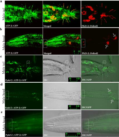
ATP-2 localizes to both cilia and mitochondria of male-specific neurons. (a and b) Confocal micrographs of Patp-2::ATP-2::GFP (left) and Ppkd-2::PKD-2::DsRed2 (right) double-labeled males (merge in middle). Adult male tail (a) and adult male head (b). PKD-2 and ATP-2 localize to cilia in the male-specific neurons RnBs, HOB, and CEMs. (c-e) Confocal epifluorescence micrographs (left), differential interference contrast (DIC) (middle), and epifluorescence/DIC images (right) of males expressing Ppkd-2::ATP-2::GFP. ATP-2 expression was restricted to male-specific neurons by using a 1.3-kb pkd-2 promoter. (c) ATP-2 localizes to ray cilia (dashed box) and mitochondria. (d and e) ATP-2 localizes to both mitochondria and cilia of CEMs. Hollowed arrows indicate CEM neuronal cell bodies. (e) Higher magnification of the nose region showing ciliary localization of ATP-2. Dashed rectangular boxes show the ciliary zone of the CEM neurons. Labeled bars indicate length in micrometers.