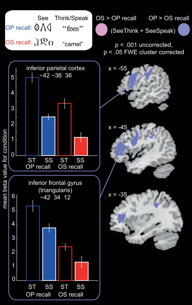
Figure 9.

Brain regions showing differential activity when recalling pronunciations (OP recall) versus meanings (OS recall) of artificial orthographies, in MRI Scan 1, prior to behavioral training. Left hemisphere slices show the results of paired t tests between these two tasks (pink: OS > OP, light blue: OP > OS), collapsed across see-think (ST) and see-speak (SS) trials. Note that no brain regions showed greater activity when recalling meanings than pronunciations. Whole-brain activations are presented at p < .001 voxelwise uncorrected and p < .05 FWE cluster-corrected for 18 participants. Plots show activation for see-think and see-speak trials for OP and OS recall at representative peak voxels that showed greater activity for recalling pronunciations (OP) than meanings (OS).
