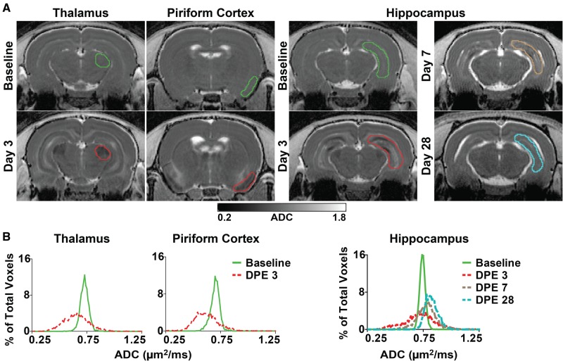
FIG. 4.
Quantification of brain damage using apparent diffusion coefficient (ADC). A, Representative, parametric maps of ADC at the level of the thalamus, piriform cortex, and hippocampus at baseline and at varying days postexposure (DPE). Colored outlines indicate slices through volumes of interest (VOIs), delineated along anatomic structures, whose pixel data contributed to the frequency distributions illustrated below. B, Frequency distributions of pixel-wise ADC values extracted from representative VOIs of the thalamus (left), piriform cortex (middle), and hippocampus (right) at baseline and 3, 7, and 28 DPE demonstrate both pronounced enhancement and restriction of diffusion within affected regions, resulting in a general broadening of the ADC distribution following acute DFP intoxication.

