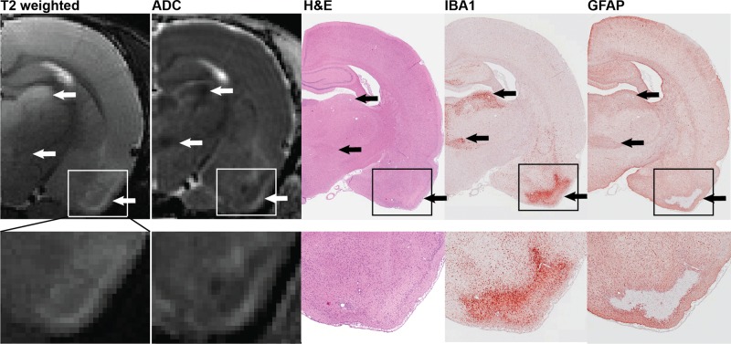
FIG. 5.
Spatial registration of lesions detected by T2 weighted images (T2w) and parametric maps of the apparent diffusion coefficient (ADC) with areas of severe neuronal necrosis, microgliosis, and astrocytic activation, as assessed by H&E staining, IBA1 immunoreactivity, and GFAP immunoreactivity, respectively. Arrows denote lesions in the dorsolateral thalamus (top), ventromedial thalamus (middle) and piriform cortex (bottom). Micrograph magnifications: hemisphere, ×0.8; piriform cortex inset, ×2.

