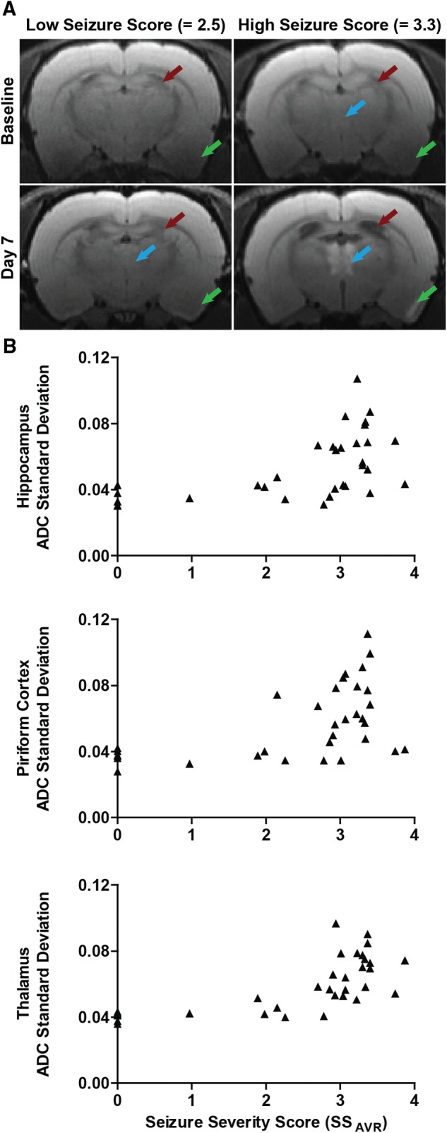
FIG. 6.

Brain damage assessed by MRI is positively correlated with seizure severity. A, Diffusion-weighted MR images of DFP-intoxicated animals with differing average seizure severity scores (SSAVR). Purple arrows identify hyperintensity in the thalamus; green arrows, hyperintensity in the piriform cortex; red arrows, expansion of the lateral ventricles. The lack of significant brain lesions in the animal with the low seizure score was confirmed by histopathological examination. B, Seizure severity, defined as the average seizure score over the first 4 h post exposure, is significantly correlated with tissue injury in the hippocampus (rs = 0.67; P < 0.001), piriform cortex (rs = 0.55; P < 0.001) and thalamus (rs = 0.74; P < 0.001), as assessed by the standard deviation of the apparent diffusion coefficient (ADC).
