
Figure 8.
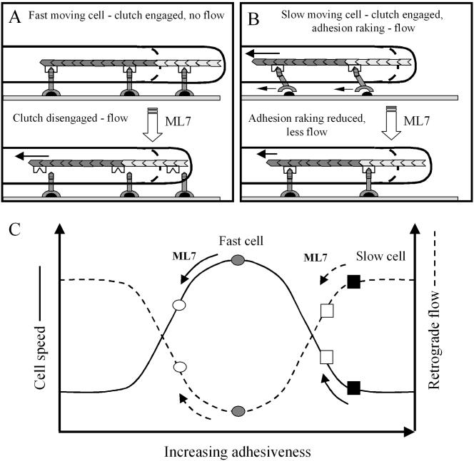
Diagrams of a protruding lamellipodium illustrating how retrograde actin flow can result from either (A) slippage between the cytoskeleton (stippled chevrons) and adhesion receptors (inverted Y-shaped symbols) or (B) raking between these receptors and their substratum bound ligands (black semicircles; B). ML7 treatment of fast-moving cells is proposed to disengage the molecular clutch (inverted M-shaped symbol) from the intracellular domain (horizontal striped region) of adhesion receptors. As a result retrograde flow increases and less newly polymerized actin (light stippled chevrons) can contribute to protrusion. (B) Raking between adhesion receptors (small, left pointing, arrows) and substratum bound ligands is shown occurring in a slow-moving cell, due to the generation of relatively large contractile forces (long arrow). In addition large contractile forces reduce the amount of actin polymerization that can contribute to protrusion. A ML7-induced reduction in contractility can reduce adhesion raking and associated retrograde flow, while increasing the rate of protrusion. (C) Hypothetical relationship between retrograde flow, cell speed and adhesiveness, that can account for the differential effects of ML7 on fast- and slow-moving cells. Cell speed (solid line) is shown to exhibit a biphasic relationship with respect to adhesiveness, which is also inversely related to retrograde flow. Rapid cell movement is shown (gray circle) to occur at an optimum level of adhesiveness, where retrograde flow is minimal. After ML7 treatment cell speed is reduced (○) and retrograde flow increases. Slow cell movement (▪) is shown to occur when adhesiveness is above optimum levels and retrograde actin flow is high. After ML7 treatment adhesiveness is reduced closer to the optimum and retrograde flow is decreased (□).