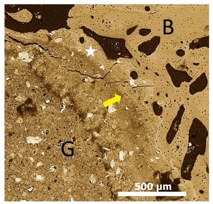
Figure 1.
Scanning electron microscope image showing calcium phosphate graft material after 12 weeks osteointegrated with bone and the osteoconduction of bone tissue around the graft material. Graft-Bone interface (Yellow arrow); existing bone (B); graft material (G); Creeping bone substitution/osteoconduction (White star).

