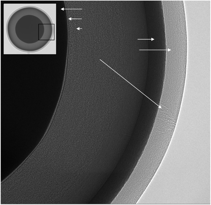
Figure 7.
2D Phase-contrast X-ray image of a fuel pellet consisting of layers of pyrolitic carbon and silicon carbide on a zirconia core. In the main image a crack can be seen, highlighted by phase-contrast, in the outer pyrolitic carbon layer. The inset is a lower magnification image providing an overview of the whole sample, approximately 800 μm in diameter.

