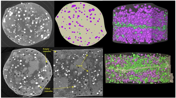
Figure 11.
Upper row; a tomographic cross section, a segmented cross section and a 3D rendered view of the seven-day healed sample showing full capsules as purple and empty ones as green (sample diameter 2 mm). Note the band of empty capsules around the crack. Lower row; two tomographic sections in different orientations and a 3D rendered view of the three-month healed sample. Note the much higher proportion of empty capsules in regions away from the crack relative to the seven-day healed sample.

