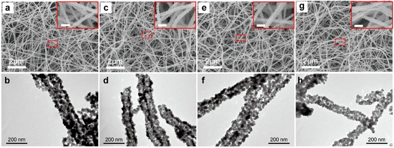
Figure 1.
Scanning electron microscope images (a,c,e,g) and transmission electron microscope images (b,d,f,h) images of (a,b) pristine SnO2 porous nanofibers as well as (c,d) 1.0 wt %, (e,f) 4.0 wt %, (g,h) 8.0 wt % Li+-doped SnO2 porous nanofibers prepared by electrospinning and post-calcination at 600 °C in air for 5 h. The insets are high-resolution SEM images (Scale bar: 200 nm). The pristine SnO2 porous nanofibers and Li+-doped SnO2 porous nanofibers showed a porous structure. The Li+-doped SnO2 porous nanofibers can be controlled by changing the ratio of lithium chloride hydrate in precursors.

