
Figure 1.
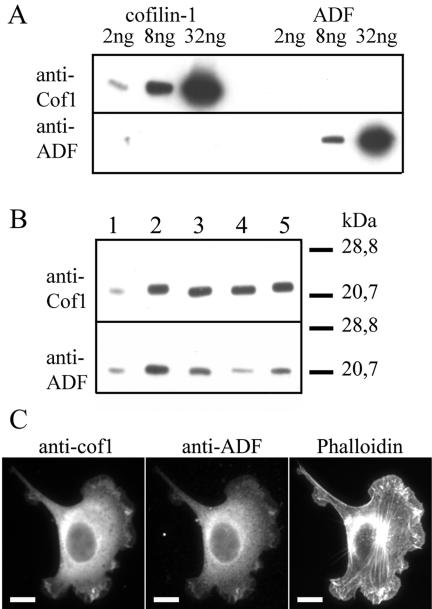
Expression levels and subcellular localizations of cofilin-1 and ADF. (A) Western blot assay demonstrating the specificities of the anti-ADF and anti-cofilin-1 antibodies. Purified recombinant cofilin-1 (lane 1, 2 ng; lane 2, 8 ng; lane 3, 32 ng) and ADF (lane 4, 2 ng; lane 5, 8 ng; lane 6, 32 ng) were visualized on Western blots by using anti-cofilin-1 (top) and anti-ADF (bottom) antibodies. (B) The levels of cofilin-1 and ADF in NIH 3T3, B16F1, and Neuro 2A cells were compared with known concentrations of cofilin-1 (top) and ADF (bottom). Lane 1, 30 ng of cofilin-1 (top) and 7.5 ng of ADF (bottom); lane 2, 80 ng of cofilin-1 (top) and 20 ng of ADF (bottom); lane 3, 10 μg of NIH 3T3 extract; lane 4, 10 μg of B16F1 extract; lane 5, 10 μg of Neuro 2A extract. Cofilin-1 is expressed approximately in sixfold (NIH 3T3), 11-fold (B16F1), and sevenfold (Neuro 2A) higher molar amounts than ADF. (C) Cofilin-1, ADF, and F-actin were visualized by immunofluorescence in NIH 3T3 cells. Cofilin-1 and ADF show similar subcellular localizations and are concentrated in F-actin-rich ruffles. Bar, 10 μm.