Figure 10.

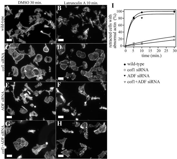
Actin filament depolymerization rates in wild-type and ADF/cofilin-1 knockdown cells. Filamentous actin was visualized by phalloidin staining in wild-type (A and B), cofilin-1 knockdown (C and D), ADF knockdown (E and F), and cofilin-1/ADF knockdown (G and H) B16F1 cells after the addition 2 μM latrunculin-A. Time points 30 min after DMSO addition (control) (A, C, E, and G) and 10 min after addition of 2 μM latrunculin-A (B, D, F, and H) are shown. (I) Percentage amount of retracted cells without clear stress fibers and with abnormal F-actin aggregates were counted (n = 50 cells) at time points 5, 10, and 30 min after latrunculin-A addition. The actin filament structures were rapidly disrupted in wild-type cells (black squares). In contrast, stress fibers disappeared much more slowly in cofilin-1 (open squares) and cofilin-1/ADF (open triangles) knockdown cells. The ADF knockdown cells (black triangles) lost their actin filament structures almost as quickly as the wild-type cells. Bars, 50 μm.
