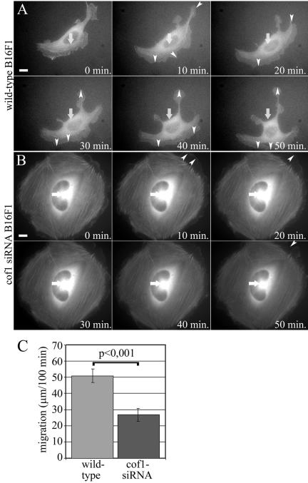
Figure 7.

Live cell analysis of wild-type and cofilin-1 knockdown B16F1 cell migration. Wild-type B16F1 cells expressing GFP-actin (A and Supplementary Video 3) displayed fast actin dynamics in the lamellipodia and directional cell motility. Cofilin-1 knockdown cells (B and Supplementary Video 4) were unable to migrate but were still capable of slowly extending and retracting their lamellipodia. White arrows indicate the locations of the nuclei in the first frame. White arrowheads indicate largest protrusions and retractions. Bars, 10 μm. (C) Migration of 35 wild-type and 25 cofilin-1 knockdown B16F1 cells were monitored for 100 min, and the positions of the nuclei was tracked every 20 min. The average motility distances of wild-type cells are 51.0 μm and cofilin-1 knockdown cells 26.9 μm. SEMs and statistical significance of the data are indicated in the graph.
