
Figure 5.
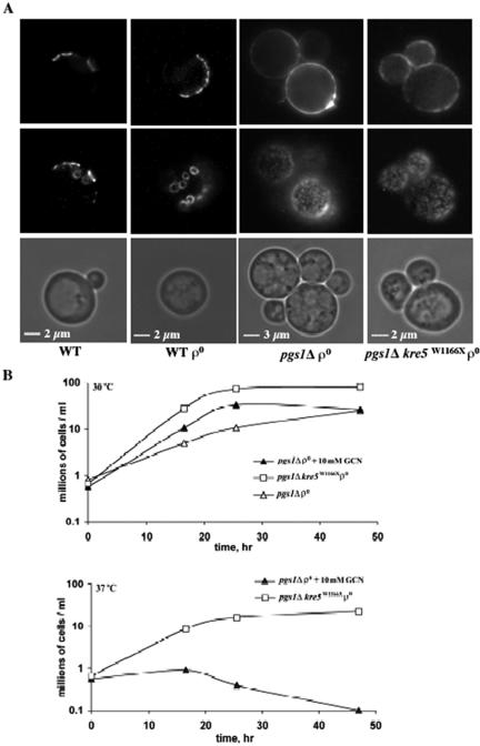
Increased chitin deposition in pgs1Δ and the suppressor mutant. (A) Cells from wild-type (FGY3), ρ0, pgs1Δ (QZY24B), and the suppressor (QZY11A) were grown in YPD to early stationary phase. Chitin was visualized by staining with Oregon Green 488 as described in Materials and Methods. Chitin distribution was visualized by focusing on two planes. (B) Cells from pgs1Δ (QZY24B) and the suppressor mutant (QZY11A) were grown in YPD in the presence or absence of 10 mM glucosamine at the indicated temperatures. Viable cells were determined by serial dilution and plating.