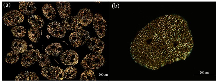
Figure 13.
Transverse section image of fiber bundles taken fromHD luffa sponge and observed by polarized-light. (a) is the transverse section image in low-magnification times, and (b) is in a high magnification times. (the noticeable dark region is the area occupied by non-fiber including cavity, vessels, and phloem tissue).

