Figure 6.

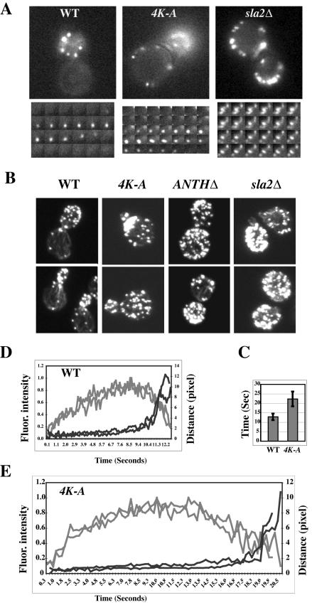
PtdIns(4,5)P2 binding is required for the normal turnover of actin cortical patches. (A) Time-lapse videomicroscopy of wild-type cells or sla2 mutants expressing Abp1-GFP. Bottom panels show selected frames of single patches at 1-s intervals. (B) Actin organization is defective in sla2 mutants. Wild-type, sla2 4K-A, sla2 ANTHΔ, and sla2Δ strains were grown on YPD to log phase at 25°C. Cells were harvested, fixed, stained with rhodamine-phalloidin, and observed by confocal laser microscopy. (C) Lifetime for Abp1 patches in wild-type or sla2 4K-A cells ± SD; n = 20. (D and E) Correlation of the formation of Abp1p patches with their movement in wild-type or sla2 4K-A mutant cells. Fluorescence intensity and distance were measured from the site of patch formation for Abp1-GFP patches over time. Each curve represents data from one patch. Fluorescence intensity over time was corrected for photobleaching. Data for two patches are shown in D and E. Gray lines represent fluorescence intensity, and black lines represent distance.
