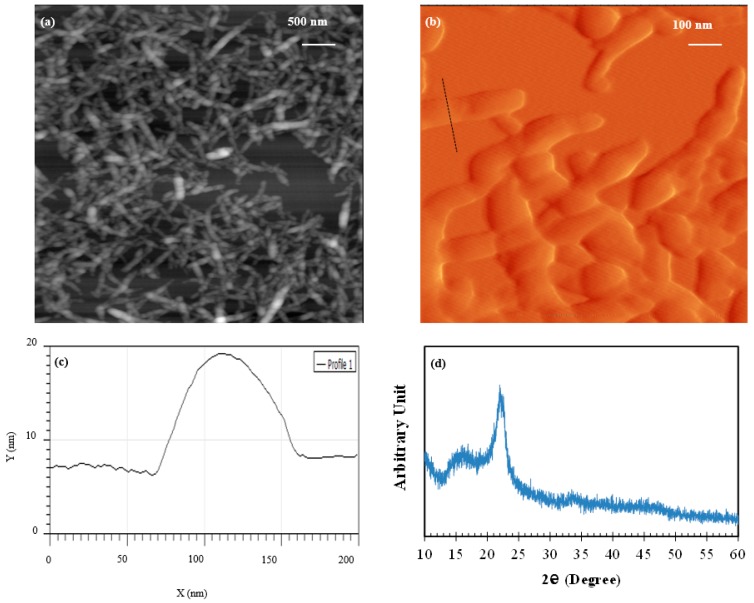
Figure 1.
(a,b) Height and error atomic force microscopy (AFM) images, respectively, of cellulose nanocrystals (CNC) taken at different magnifications; (c) Line profile of a CNC nanofiber along the line marked with a dashed line in Figure 1b; (d) XRD spectrum of CNC.

