
Figure 5.
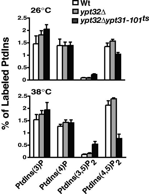
Phosphoinositide levels in Wt, ypt32Δ, and ypt32Δypt31-101ts double mutant cells. Cells were incubated at the temperature indicated for 10 min and labeled with myo-[2-3H]inositol for 45 min, and phosphoinositides were analyzed as previously described in Rudge et al. (2004). The levels of each indicated phosphoinositide are expressed as a percentage of the total 3H-labeled phosphoinositides analyzed by HPLC and represent the average of two independent experiments done in duplicate (n = 4).