
Figure 6.
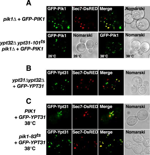
Independent localization of Pik1p and Ypt31p to the trans-Golgi network. (A) pik1Δ cells expressing GFP-Pik1p and Sec7p-DsRed were examined by fluorescence microscopy at 26°C. ypt32Δypt31-101tspik1Δ cells expressing GFP-Pik1p were grown at 26°C or shifted for 1 h at 38°C and examined by fluorescence microscopy. (B) ypt31Δ ypt32Δ cells expressing GFP-Ypt31p and Sec7p-DsRed were grown at 26°C and examined by fluorescence microscopy. (C) Wild-type (PIK1), pik1–83ts, or pik1-139ts (unpublished data) cells expressing GFP-YPT31 from a low copy plasmid and Sec7p-DsRed were grown at 26°C (unpublished data) or shifted 1 h at 38°C and examined by fluorescence microscopy.