
Figure 11.
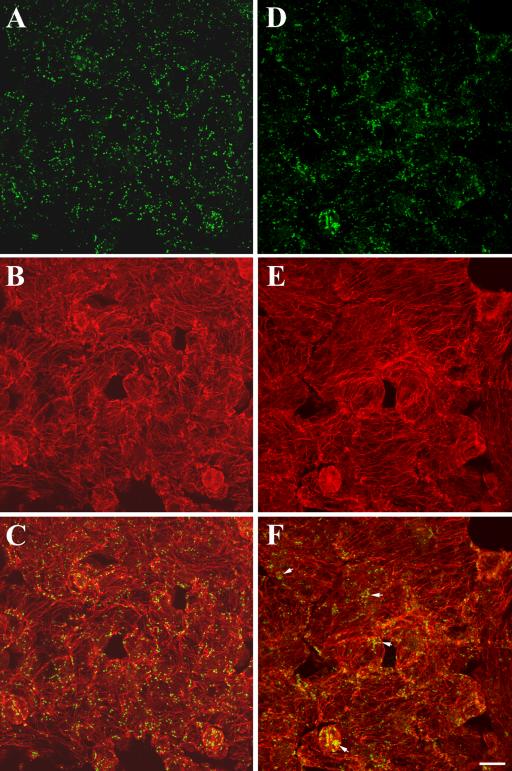
Distribution of Golgi stacks in leaf epidermal cells of wild-type and the kinesin-13a-1 mutant. (A–C) Wild-type cells. (A) Golgi stacks revealed by anti-α-mannosidase were evenly distributed in epidermal cells. (B) Cortical microtubules stained by anti-α-tubulin. (C) Merged image showing both Golgi stacks and microtubules. (D–F) kinesin-13a-1 mutant cells. (D) Golgi stacks revealed by anti-α-mannosidase. Although many Golgi stacks were still dispersed, in a number of places, Golgi stacks seemed to be more aggregated than in wild-type cells. (E) Similar cortical microtubule network was observed as in wild-type cells. (F) Merged image. Note aggregation of Golgi stacks was obvious in guard cells and other epidermal cells (arrows). Bar, 10 μm.