
Figure 9.
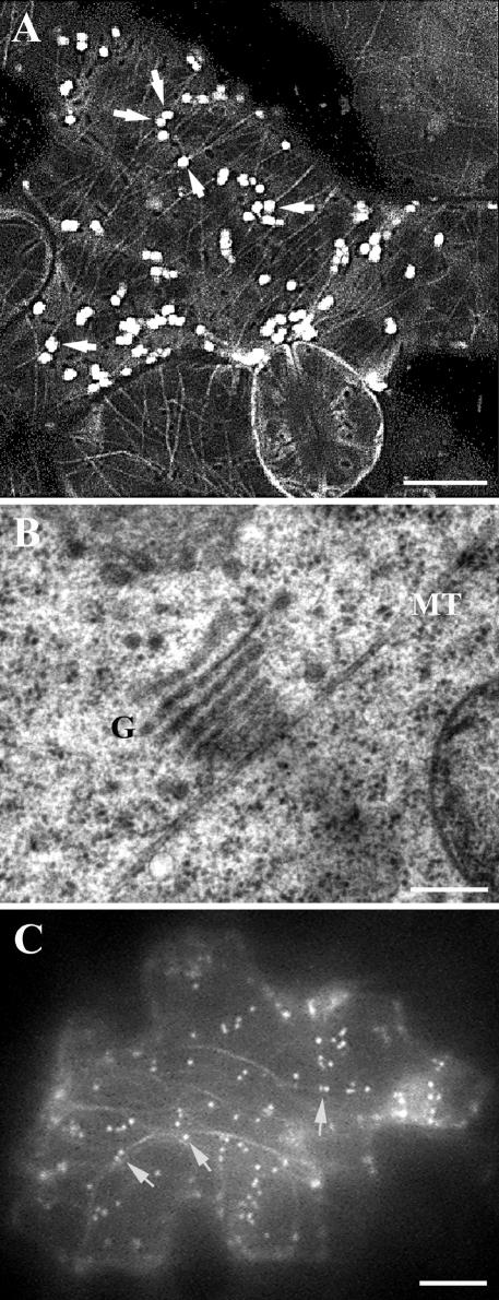
Association of Golgi stacks with microtubules and actin microfilaments. (A) Confocal image showed association of Golgi stacks labeled with Nag-GFP with cortical microtubules labeled with GFP-α6-tubulin (arrows). Note cortical microtubules were present in all cells. Bar, 10 μm. (B) Transmission electron microscopic image showed that a Golgi stack (G) was associated with a microtubule (MT). Bar, 0.2 μm. (C) Wide-field image showed association of Golgi stacks labeled with Nag-GFP and GFP-talin (arrows). Nag-GFP labeled Golgi stacks as bright particles, and GFP-talin labeled actin microfilaments as dimmer filamentous structures. Bar, 10 μm.