
Figure 5.
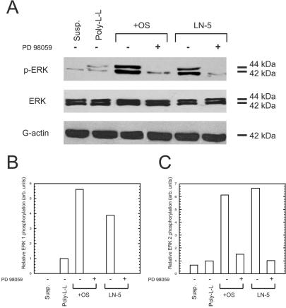
Adhesion to Ln-5 activates ERK in hMSC. (A) Cells kept in suspension (susp.), or plated for 30 min on poly-l-lysine (Poly-L-L), tissue culture plastic with OS media (+OS), or on Ln-5 were probed by immunoblot for phosphorylated ERK (top row), total ERK (middle row), or actin as a loading control (bottom row). Where indicated by a “+” symbol, 50 μM PD98059 was added to the culture conditions. (B and C) Densitometric measurements of band intensities for phospho-ERK 1 and phospho-ERK 2, respectively.