
Figure 7.
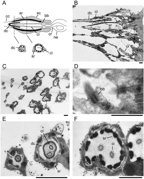
Ultrastructure of chordotonal organs in DmEB1P mutant. Electron micrographs of auditory chordotonal organs in Johnston's organs from DmEB1P mutant. All prominent ultrastructures with normal morphology are observed. Bar, 1 μm. (A) Schematic diagram of each sensory unit of the auditory chordotonal organs, modified from Dubruille et al. (2002). Top, longitudinal section. Bottom, cross sections. bb, basal body; cc, cap cell; ci, cilium; cr, ciliary rootlet; dc, dendritic cap; sc, scolopale cell; sr, scolopale rod; and ne, neuron. (B) Longitudinal section revealing a dendritic cap, cilia, scolopale rods, and ciliary rootlet. (C) Cross section of multiple chordotonal organs. More proximal cross sections of the organs are observed toward the left. (D) Higher magnification of a longitudinal section showing basal bodies. (E and F) Higher magnification of different cross sections. Dendritic caps are visible between cilia and a circle of scolopale rods near the distal ends (E) but absent from more proximal positions (F).