
Figure 2.
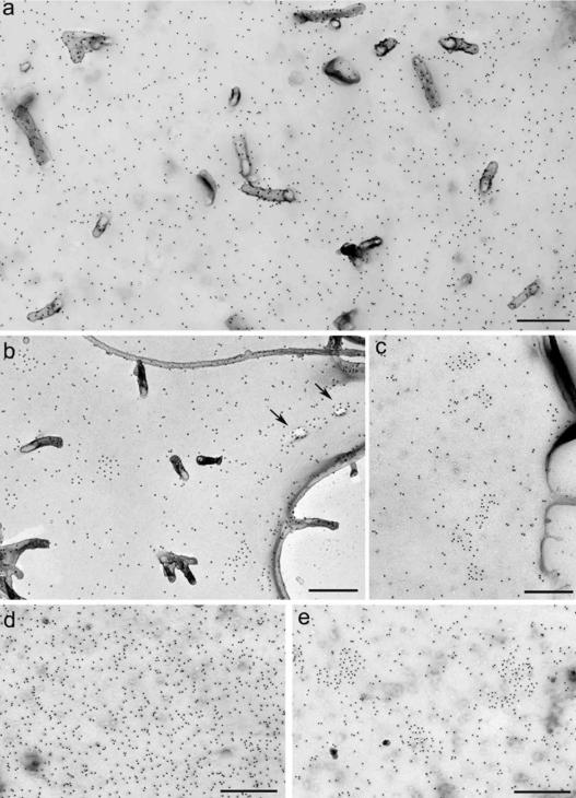
CCR5 cell surface distribution. CHO-CCR5 (a–c) and RBL-CCR5 (d and e) cells were treated in BM alone (a and d) or with CCL5 for 30 s (b) or 5 min (c and e) at 37°C before fixation. Surface CCR5 was detected by labeling with MC-5 and PAG15, and cell surface replicas were prepared as described in Materials and Methods. Agonist-treated CCR5 was occasionally seen associated with shadowed invaginations of the membrane (arrows). Bars, 500 nm.